Citation: News of Beam
Diagnostics Belarus 1999 3: 21-23.
Role of
computed tomography in diagnostics of colonic neoplasias.
Shaparov I. N.1,
Ovchinnikov V. A.2
1Grodno Regional Clinical Hospital,
2Grodno State Medical Institute, Grodno.
|
|
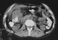
| Figure 1.
CT scan shows hepatic flexure colon carcinoma. Colon walls thickened
irregularly, surrounding fat is infiltrated, adjacent peritoneum thickened
as well. |
|
|
|
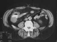
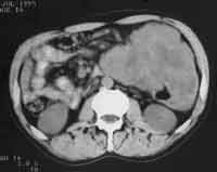
| Figure 2.
Caecum carcinoma: irregular walls thickening, surrounding fat infiltration. |
| |
|
|
|
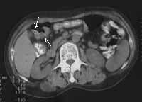
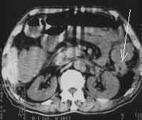
| Figure 3.
Transverse colon carcinoma: local thickening of the posterior wall
near hepatic flexure (arrows), enlarged regional lymph nodes. |
|
|
|
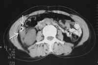
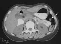
| Figure 4. Ascending colon carcinoma: irregular walls thickening (arrows)
with renal fascia involvement. |
|
|
|
| Figure 5. Descending colon carcinoma (arrow): irregular walls
thickening, surrounding fat infiltration. |
|
|
|
| Figure 6. CT scan shows huge tumour surrounding descending
colon. |
|
|
|
| Figure 7. Stalk polyp (arrow) is well depicted by CT scan
at the background of gas in the lumen of colon spleen flexure. |
|