Citation: News of Beam Diagnostics Belarus
2000 1: 8-11.
Chronic visceral hemodinamics disorders.
Filippovich N. S.
Belarussian Medical Academy of Postgraduate Education, Minsk.
|
|
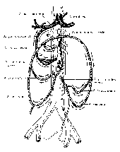
| Figure 1. Schema depicts collateral circulation pathways in
abdominal angina syndrome. |
|
|
|
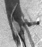
| Figure 2. Celiac trunk stenosis in atherosclerosis. Lateral
angiogram. |
|
|
|
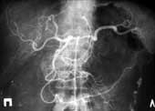
| Figure 3. Celiac trunk and spleen artery stenosis in atherosclerosis.
Selective celiac angiography. Poststenotic dilatation of common hepatic
artery. |
|
|
|
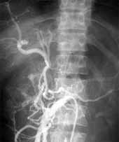
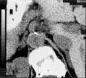
| Figure 4. Selective superior mesenteric artery angiography.
Celiac trunk and hepatic artery contrasted retrogradely via “small arc”. |
|
|
|
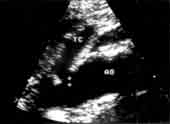
Figure 5. Ultrasound scan shows atheroma in celiac trunk orifice.
TC - celiac trunk,
АО - aorta. |
|
|
|
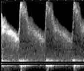
| Figure 6. Blood flow spectrogram demonstrates turbulent flow
at the site of celiac trunk stenosis. |
|
|
|
| Figure 7. Abdominal vessels atherosclerosis. CT scan shows calcinats
in abdominal aorta wall just near celiac trunk orifice. |
|